

- Home
- Companies
- Regent Instruments Inc.
- Software
- WinCELL - Wood Cell Image Analysis ...
WinCELL - Wood Cell Image Analysis Software
WinCELL is an image analysis system specifically designed for wood cells analysis. It can quantify the changes in wood structure over annual rings.
Anatomical wood cell analysis is an alternative to wood density analysis with x-rays (as done with WinDENDRO). Wood density, color, mechanical and chemical properties are in effect related to wood structure which in turns is related to climate. By measuring the radial cells (tracheids) size, distribution and their proportion to walls, wood quality can be assessed.
WinCELL measures wood cell morphology on thin wood slices cut with a microtome or, for larger cells like earlywood vessels of deciduous species, directly on wood surfaces*. Wood cells morphological data can be measured per annual ring in one or more images per ring with the aid of XLCell, a companion program for data post-processing and visualization.
Image analysers not made specifically for wood cells measurement are not usually able to produce data suited for dendrochronology studies. These systems lack some knowledge about annual tree rings formation and the structure of their cells (to compute the ring width for example). WinCELL has this kind of know-how built-in. It knows, for example, that a wall between two adjacent cells must be split in two to compute the cells length and earlywood or ring width. Its versatile settings allow to analyse different wood species (vessels of deciduous and radial row of conifer tracheids). It supports different automatic and interactive analysis modes. The latter allows you to select rows of cells to analyse them in a way that mimics the traditional trachedoid** method. WinCELL handles incomplete cells, those truncated by image boundary, so that they have no effects on the average cell measurements.
*With proper image acquisition device and sample preparation.
**Tracheidograms are curves of radial cell size variations in function of position in an annual ring.
Image Acquisition
Recent developments in high-quality affordable digital cameras have made anatomical wood cell analysis more accessible than ever.
Wood cell analysis is traditionally done on thin wood slices cut with a microtome. To acquire images of such samples, a microscope with a receptacle tube for a camera attachment and a camera adapter are required. Staining is sometime used to enhance lumen-wall contrasts when acquiring images in translucent light. This is the ideal setup to acquire very high resolution and precise images but the field of view is usually limited so that it is difficult or impossible to view entire rings. Such images can be analysed and their data merged afterward in XLCell to do cells analysis on a ring basis.
Alternative sample`s preparation methods and imaging over larger areas also exists since some time but none is universally used. These are typically based on a scanner or a camera and macro lens and proper lighting. The difficulties with this approach are: to acquire images with good enough contrasts between lumen and walls, to do so without damaging them and to get enough resolution to accurately identify and analyse them. Their advantage is to allow to acquire images of a few annual rings per image, making their analyses easier.
WinCELL can analyse images acquired with scanners or digital cameras. The camera we sell is of scientific quality grade and have a standard C-mount thread which allows it to be installed on a microscope using an adapter (C-Mount adapter, not included). This camera can also be placed directly above the wood sample to acquire images with optional lenses. It is interfaced to computer via a fast USB connection. If you plan to use a scanner please inquire about the conditions before purchasing. WinCELL is TWAIN compatible, meaning it can acquire images from a few camera or scanner models.
WinCELL uses the concept of analyzed region in order to reject incomplete cells. Cells touching the image boundary or located outside the analyzed region are not considered when computing average cell measurements (area, length and width). The color used to draw a cell indicates its classification: partially or completely outside or inside the analysed region, rejected by operator, debris, cell type (cell, vessel or parenchyma).
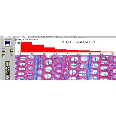
Measurements data are available interactively during the analysis and in text files that can be read by many software programs. These files are easily opened and visualized in spreadsheet style programs like Microsoft Excel. You can also click a cell to display its morphological measurements data. The cells distribution histogram, visible during the analysis or after in XLCell, also present a global view of the cell structure parameters. The cells distribution histogram displays the number of cells in function of area, length or width and for the Pro version the area in function of color.
Image edition allows to compensate for defects or poor contrast. Images can be edited with any color. It is easy to select a color present in the image and edit with it. A pen (to draw lines) and a lasso tool (to fill outlined regions) are provided for edition.
Miscellaneous Features
- WinCELL can analyse grey levels or color images (our cameras produce both kinds). The Pro version can do more analyses on color images. It can display and analyse one of its three color channels, use the color content to better classify the pixels into lumen and wall or quantify area in function of color.
- Calibrations are easy to perform on targets sold by microscope manufacturers. Different target models are supported.
- Debris (defects or non cells objects) can be automatically filtered out by morphological features (area, length, width, form, length to width ratio), color (Pro version) or by editing the images.
- Original images acquired from WinCELL, analysed or not can be saved in standard tiff or bmp files for opening in other application programs. Images saved with their analysis (in the same file) are automatically reanalysed when reopened in WinCELL (useful to validate or modify a previously done analysis).
- Batch processing is provided to analyse a series of images without operator supervision. This analysis mode works only for analyses that can be done automatically (non-interactively).
- It is possible to store the analysis settings in configuration files for retrieving and reusing at a later time.
- You can choose which data are saved.
- WinCELL can also be used as a general area meter (to measure leaf area for example) or a morphology analyser for other objects by modifying its default settings.
- Like all Regent`s products, WinCELL is a stand-alone program with all the built-in necessary functionality. It does not require an additional complex image analysis program or user programming skills as in some other cell analysis programs